
The Physics behind an ECG
It's not measuring your heart. It's measuring which way a wave is pointing.

By the end of this, you’ll look at an electrocardiogram and know exactly what you’re seeing. From the medical side, and from the physicist’s side.
But before we go into the heart, I want to walk back through a couple of physics ideas. Nothing heavy. Most of this you already met around grade eight.
You’ve probably seen the force between two charges. We say a charge q₁ feels a force from a charge q₂:
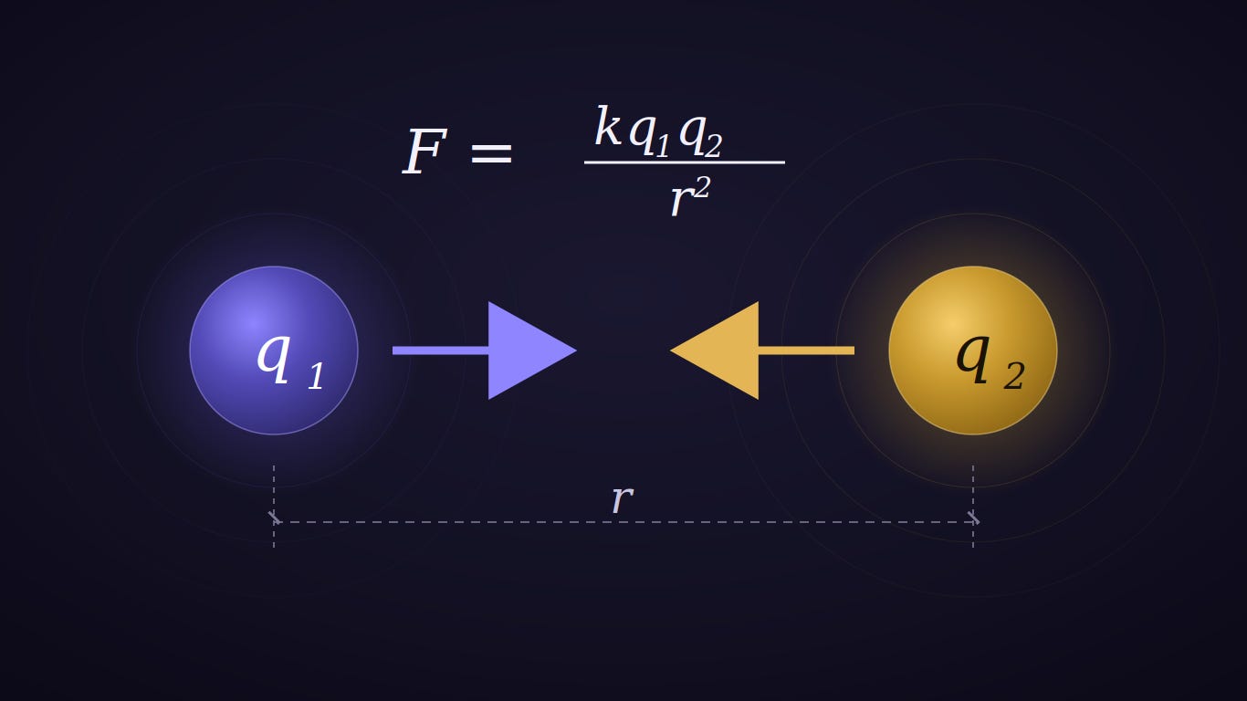
Simple enough. The force grows with the size of the charges, which makes sense. Bigger charges push or pull harder. And it falls off with distance, also no surprise. The farther apart they are, the weaker the force. Notice the distance is squared, so moving twice as far away doesn’t halve the force, it quarters it.
That’s the whole thing. Grade eight. Most of you knew it before you opened this.
But it’s not a very elegant way to think once you have more than two charges. Tracking every pair pulling on every other pair gets ugly fast. So physicists do something cleaner. Instead of asking “what force does q₂ feel from q₁,” we look at one charge and say: this charge fills the space around it with a field.
A single charge sets up a sphere of influence, attraction or repulsion depending on its sign, getting weaker the farther out you go. Like the image below.
E = kq1/r^2
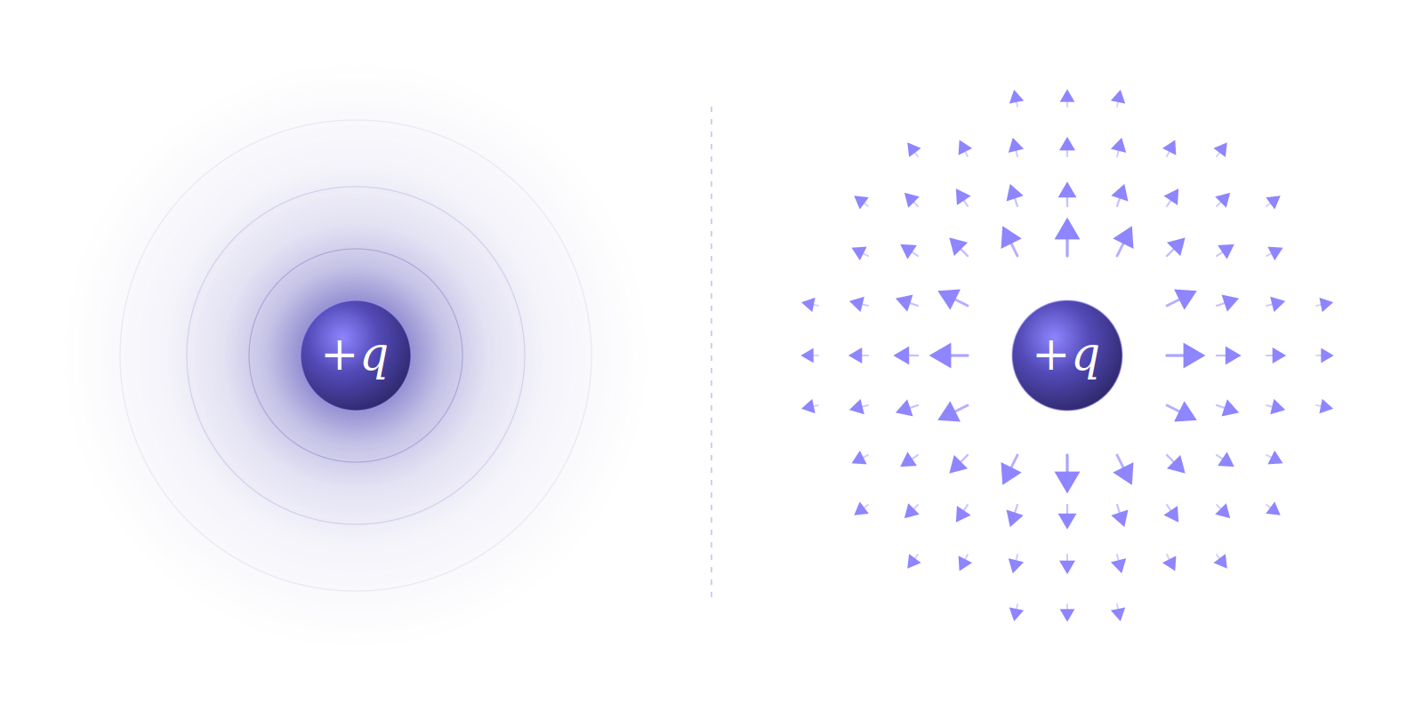
Notice arrows are outside since charge is positive( this is just a model it could’ve been to the inside).
Any other charge that wanders into that field feels a push or a pull, set by the same formula as before.
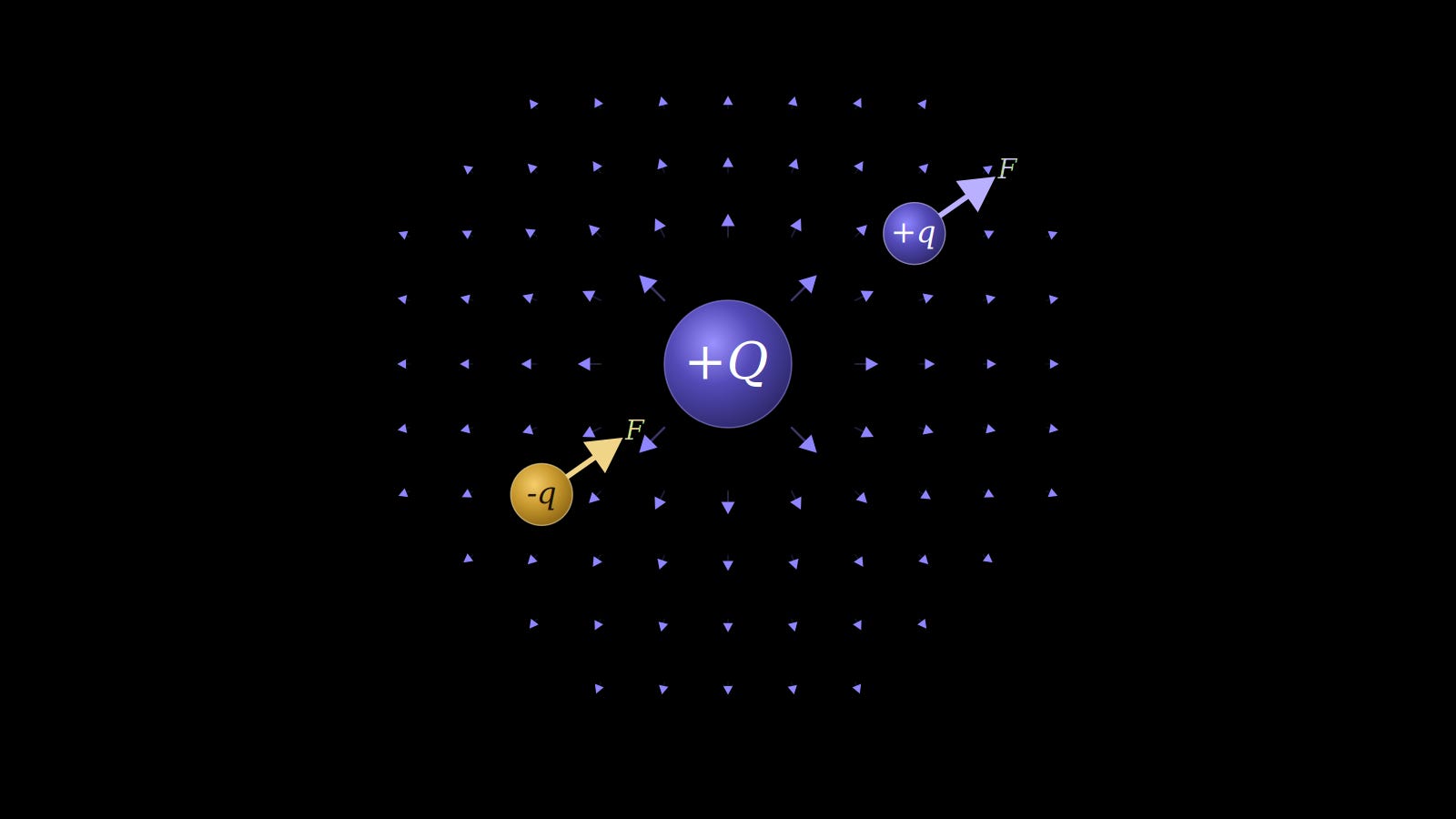
F = q2.E look how elegant the vectors now make positive and negative charges. Now this article shouldn’t be about the elegant of this model but I get carried away easily. We’ll reach the heart soon so stay with me.
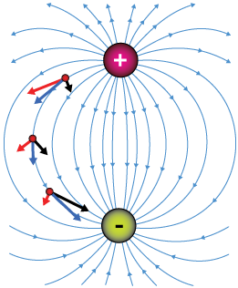
Change the shape of the source and the field changes shape with it. Two opposite charges next to each other, known as a dipole, makes this shape.
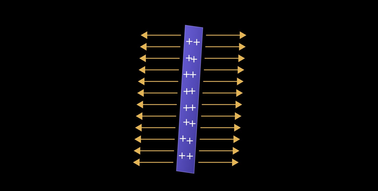
A flat sheet of charge creates this.
Here’s why I’m dragging you through this. When a physicist wants to understand anything electrical, this is the move. We don’t chase individual charges. We look at the field they make. The field is the thing we measure.
Hold also onto that word. Dipole. And hold onto that idea, that we read electricity by reading its field. Because in a few minutes you’re going to find out that your heart is a dipole, and the electrocardiogram is nothing more than a physicist standing at a distance, measuring its field.
Now, when you hear about electricity, you never hear about any of this, and for a good reason. It gets complicated fast. What you hear about instead is voltage. And from everything we just built, voltage is suddenly easy to define.
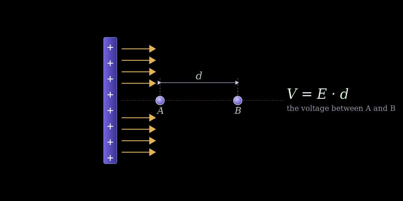
It’s simply a scalar quantity of the field that assigns a “potential” to each position. And I know physics picks intimidating words sometimes, but this one is exactly as plain as it sounds. A potential on its own means nothing. What we actually care about is the potential difference, which just tells you how much the field’s effect changes between two positions. How do you get it? For a uniform field, it’s distance times the field. Nothing fancier.
And here’s the part I find beautiful. The potential energy of a charge sitting in that field is just the charge times V. Look at the pattern. F and E related the same way E and V do, and that’s not a coincidence. For those of you who know some calculus: E is what you get when you differentiate V across distance, the same way force comes from differentiating energy. Integrate back the other way and V is the field summed over a distance.
Sorry for the long road. But we landed somewhere genuinely clean. Voltage difference is, in a real sense, a measure of how much electricity a position carries. And that is exactly what you’ll see on an ECG.
So, finally, let’s go to the heart.
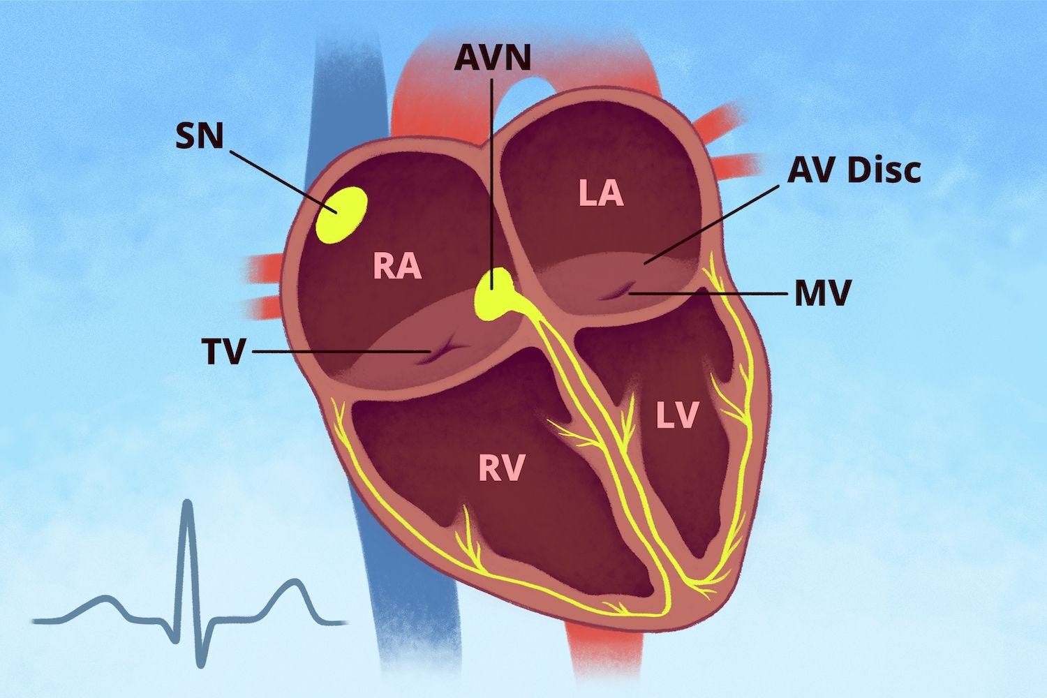
This is a cross-section of the heart, drawn as if you’re looking at someone standing in front of you. That detail matters, because it flips everything: their right side is on your left. So on the left of the image you’re seeing the right atrium (RA) and right ventricle (RV), and on the right of the image, the left atrium (LA) and left ventricle (LV). Atria on top, ventricles below.
Now a little electrophysiology.
The heart beats somewhere around 60 to 100 times a minute at rest. Here’s the first surprising thing: nothing outside the heart is telling it to. The beat starts inside, at a small patch of tissue in the wall of the right atrium called the sinoatrial node, the SA node. And the SA node is not made of nerves. It’s specialized muscle, cardiac cells that have given up squeezing in order to do one thing instead: fire, rhythmically, on their own. Cut every nerve to the heart and it keeps going. Your brain can lean on the SA node to speed it up or slow it down, but it never starts the beat. The heart paces itself.
When one of these cells fires, what we medically call depolarizing, it doesn’t keep the charge to itself. It dumps it into the cell touching it, which depolarizes in turn and passes it on. The signal spreads the way a rumor does, cell to cell, no wire required. The exact description of this spreading is biochemical where each cell has a sensor that fires when the cell beside it has changes electricity (voltage gated). And it gets electrically excited by movement of atoms inside of it (Sodium, calcium and potassium). So now we have charged sections of the heart creating an electric field creating a potential as we explained previously
That spreading wave follows a set path, the yellow line in the image. It starts at the SA node and sweeps across both atria, so the RA and LA contract first, squeezing blood down into the ventricles. Then it reaches a second relay station between the atria and ventricles, the atrioventricular node (AV node). From there it runs down a fast cable through the wall dividing the two ventricles, and fans out through fibers that carry it up the ventricle walls from the bottom.
That fast cable matters more than it sounds. The signal reaches the bottom of the ventricles first, almost simultaneously, so the whole lower mass fires in one near-instant burst rather than slowly creeping cell to cell. That coordination is the point. The ventricles are the big muscular pumping chambers, and you want all that muscle contracting together, in one hard coordinated grip, not in a slow ripple. The fast wiring is what buys that synchrony. It’s the difference between a fist clenching all at once and fingers curling one at a time.
That’s the wave. Hold onto it, because everything on the ECG is just this wave, seen from a distance.
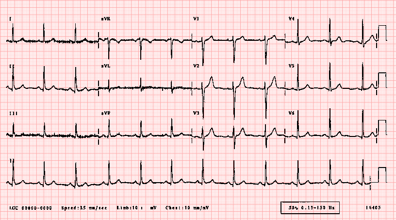
So how do we actually watch this wave? We do something clever. We put electrodes on the skin, ten of them, and from those ten we manufacture twelve different views of the heart.
That gap between ten and twelve is the first trick, and it’s worth slowing down on. An electrode is not a view. An electrode is just one sticker reading the electrical altitude at one spot on your body. On its own it tells you nothing, remember, a single potential is meaningless. You only get a signal when you take the difference between two of them. So a “lead,” in ECG language, is not a wire or a sticker. A lead is a pair: one electrode you treat as the low end, one as the high end, and the lead is the voltage running between them as the heart’s wave sweeps past.
I’ll be honest about something that bothered me when I first learned this. “The voltage running between two electrodes” is technically right, but it feels hollow. Voltage is a number. A number doesn’t move. And the heart’s signal is the most directional thing in the body, a wave sweeping across the muscle in a specific direction. How does a bare number capture a direction?
It doesn’t, on its own. But remember where voltage came from. Way back at the start, voltage wasn’t the fundamental thing. The field was. Voltage is just the field’s scalar shadow, the field measured across a distance: V = E.d. The number always had a field hiding inside it.
So here’s the honest version of what an electrode does. It can only read one thing, the potential at its spot, a scalar, a single altitude. And a lead reports the difference between two of them. That’s voltage, full stop, that’s all the machine ever measures.
But because of V = E·d, that voltage difference is really the field projected onto the line between the two stickers. It’s a dot product: the heart’s field vector, dotted with the line connecting the two electrodes. And a dot product has a beautiful property baked in. It depends on the angle between the two. When they’re perpendicular, it’s zero. When they point the same way, it’s large and positive. When they point opposite ways, it’s large and negative. The angle is everything.
So when the heart’s wave sweeps along the lead’s line, pointing the same way, the projection is large and the voltage swings hard and positive. When the wave sweeps along that line but the other way, the voltage swings hard the other direction, negative. And when the wave moves sideways, perpendicular to the line, the projection collapses to nearly zero and the voltage barely flickers. The electrode is measuring a scalar, a single number, but what that number is tracking is the field’s direction in space, read off as an angle.
That’s why the field picture from the intro wasn’t a detour. The number is what we measure. The field is what’s actually moving. And a lead is the one quietly translating the other, beat after beat.
Pick a different pair, and you’ve built a different lead. Same ten stickers, recombined, twelve times over. Twelve cameras, ten tripods. The electrocardiogram machine is really smart, it can pick electrodes and check the potential difference between them creating a lead
And here’s the way to think about each one. A lead is an eye. It sits at a fixed spot and stares at the heart along one particular line, the line drawn between its two electrodes. It can only see motion along that line. A wave coming straight toward the eye looks like one thing; the same wave moving sideways across its gaze looks like almost nothing at all.
Keep reading with a 7-day free trial
Subscribe to Physics Gene to keep reading this post and get 7 days of free access to the full post archives.